
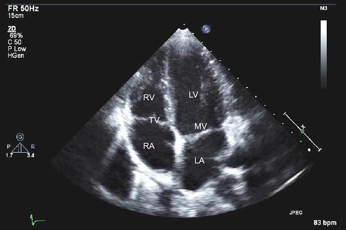
2D Echo
It is a non-invasive heart investigation that creates images of the sections of the heart using sound vibrations. It presents the various parts of the heart as in pictures so that it becomes easy to check if there is any damage or blockages, and blood flow rate.
an echocardiogram is a noninvasive (the skin is not pierced) procedure used to assess the heart's function and structures. During the procedure, a transducer (like a microphone) sends out sound waves at a frequency too high to be heard. When the transducer is placed on the chest at certain locations and angles, the sound waves move through the skin and other body tissues to the heart tissues, where the waves bounce or "echo" off of the heart structures. These sound waves are sent to a computer that can create moving images of the heart walls and valves.
A 2-D echo view appears cone-shaped on the monitor, and the real-time motion of the heart's structures can be observed. This enables the doctor to see the various heart structures at work and evaluate them.
TPA Empanelment
Cashless facility for nearly all major TPA providers